Primal 3D Series: Sports Injuries Knee, 2nd Edition
$30.00
“This product shows the true potential of the use of multimedia in medical education
Primal 3D Series: Sports Injuries Knee, 2nd Edition
Check it out: Primal 3D Series: Sports Injuries Knee, 2nd Edition
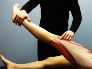
An essential reference for anyone involved in the diagnosis and treatment of knee injuries, Sports Injuries: The Knee, Second Edition, provides a complete and accurate anatomical reference in stunning 3-D and a comprehensive guide to acute and chronic overuse injuries of the knee.
The anatomy section includes a high-resolution three-dimensional computer graphic model of the complete anatomy of the knee. You can choose from a variety of views of the model, rotate at any point, and peel away layers of anatomy from skin to bone. Clicking on any features or structures within the model brings up extensive anatomy, clinical, and pathology text supported by hundreds of color diagrams, videos, and slides.
The second edition features
-better 3D graphics with new 3D views that include bursae, fascia, lymph system, and synovial membranes;
-larger rendered images so the images can be viewed in a larger format when running the software;
-new 3D animations including skeleton with muscles running, throwing, and kicking a ball;
-animations of overuse injuries; and
-new 3D illustrations of injuries to accompany clinical text.
The injuries section has relevant pathology, ligament tests, surgical treatment, conservative management, and rehabilitation of many common and less common injuries of the knee. More than 180 physiotherapy and biomechanics slides and videos in this section provide unprecedented coverage of knee injuries. The acute injuries section covers meniscal tears, articular cartilage injury, fractures around the knee, patella tendon rupture, bursitis, and reflex sympathetic dystrophy (postinjury). The overuse injuries section has information on anterior knee pain (e.g., patellofemoral joint syndrome), lateral knee pain (e.g., iliotibial band friction syndrome), medial knee pain (e.g., synovial plica), and posterior knee pain (e.g., knee joint effusion). All of these examples have related slides and video clips.
Sports Injuries: The Knee, Second Edition, also includes a quiz and test facility, the ability to import any image into your own private educational presentations, royalty free, and will work on both Windows and Macintosh.
Course instructors: Adopt Primal software for use in your class!
Teaching the intricacies of anatomy to your students has never been easier. Primal Pictures software programs allow you to illustrate anatomy to your students in remarkable new ways, and can be used in laboratory settings or in the classroom. Take advantage of special pricing on network versions for individual products or for the entire line of Primal software through an outright one-time purchase or a renewable license agreement. Several billing options are available based on the number of students in your course. These programs can be delivered via the Internet or through network configurations that can be constructed with your specific needs in mind. If you are interested in adopting this software for your class, please contact a sales representative at the phone numbers below for details!
| HK USA – Wes Osmon | (800) 747-4457 ext. 2430 |
| HK Canada | (800) 465-7301 |
| HK Europe | +44 (0) 113 255 5665 |
| HK Australia | (08) 8372-0999 |
| HK New Zealand | (09) 448-1207 |
For a complete selection of Primal Pictures software, visit www.HumanKinetics.com/Primal.
System Requirements
Windows
-Windows 98/2000/ME/XP
-Pentium processor or equivalent
-32 MB RAM
-16-bit color display
-CD-ROM drive
Mac
-G4 processor or greater
-32 MB RAM
-16-bit color display
-Mac OS 9 or greater
-CD-ROM drive


